- Home /
- University /
- Infoservice /
- Press Releases /
- Spectral microCT: new tools for X-ray imaging of microvasculature
Research
Spectral microCT: new tools for X-ray imaging of microvasculature

A study by the University of Veterinary Medicine Vienna published recently in the “Journal of Microscopy” presents a new workflow for the three-dimensional visualization and analysis of microscopic blood vessels using X-ray computed tomography (microCT) in laboratory animal models. According to the researchers, the new method is an important step towards a better understanding of smallest blood vessels and vascular disorders.
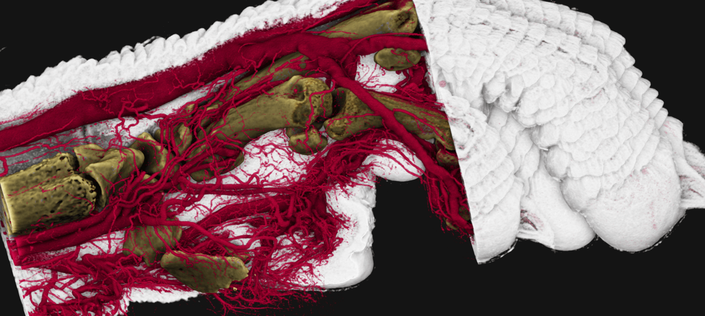
Imaging of microscopically small blood vessels using X-ray contrast agents has been established for many years and utilized by several international research groups for the quantitative analysis of blood vessels in laboratory animals. The present study introduces novel microscopic dual-energy CT (microDECT) imaging protocols that make it possible to visualize and analyze the microvasculature of organs or body regions in situ and with reference to the morphology of hard and soft tissues.
Imaging with the dual-energy CT method allows the spectral separation of blood vessels and surrounding tissue. First author of the study, Stephan Handschuh from the Imaging Core Facility at Vetmeduni: “Using dual-energy CT, we can spectrally distinguish blood vessels from bones and surrounding organs based on their X-ray properties. This allows us to generate multi-colored microCT data.”
New workflow optimizes research practice
In contrast to traditional grayscale X-ray images, multi-colored visualizations are now possible. In this approach, the visualization of organs and tissues is accomplished by a complementary X-ray contrast agent. Using these methods, microscopically small vessels can be differentiated in color from surrounding bones, organs and tissues and can therefore be displayed and analyzed in the context of the surrounding tissue. Counterstained samples can also be automatically processed into three separate image channels - skeletal tissue, vessels and stained soft tissue - offering numerous new options for data analysis.
The new workflow provides clear benefits for research practice, as Stephan Handschuh explains: “The method we have developed offers scientists new possibilities for imaging blood vessels and thus new analysis tools for studying vascular diseases.”
Better and faster analysis options in laboratory practice
The method enables new approaches for analyzing the blood vessel supply of organs. In the present study, this is demonstrated on the brain. Dual-energy CT imaging allows the brain anatomy to be included in the analysis of the vessels and thus, for example, the blood vessel volume for certain regions of the brain can be determined.
Martin Glösmann, Head of the Imaging Core Facility at Vetmeduni, explains how the new workflow opens up new possibilities as follows: “The three-dimensional representation of blood vessels in the context of the surrounding tissue is particularly suitable for recording changes to organs in genetically modified laboratory animal models quickly, quantifiably and with high throughput.”
Imaging Core Facility of the Vetmeduni
The study is a project of the Imaging Core Facility of the Vetmeduni. In addition to providing access to routine techniques, the development and establishment of new methods is one of its tasks. The working group headed by Martin Glösmann has many years of experience in the development of new X-ray contrast techniques and is one of the leading working groups in the field of microscopic dual-energy CT.
The article „In situ isotropic 3D imaging of vasculature perfusion specimens using x-ray microscopic dual-energy CT“ by Stephan Handschuh, Ursula Reichart, Stefan Kummer and Martin Glösmann was published in „Journal of Microscopy“.
Scientific article
Scientific article:
Dr. Stephan Handschuh
VetCore Facility for Research, Imaging Unit
Veterinärmedizinische Universität Wien (Vetmeduni)
stephan.handschuh@vetmeduni.ac.at